
What 3D Imaging Can Show That a Regular X-Ray Can't: How CBCT Helps Plan a Root Canal

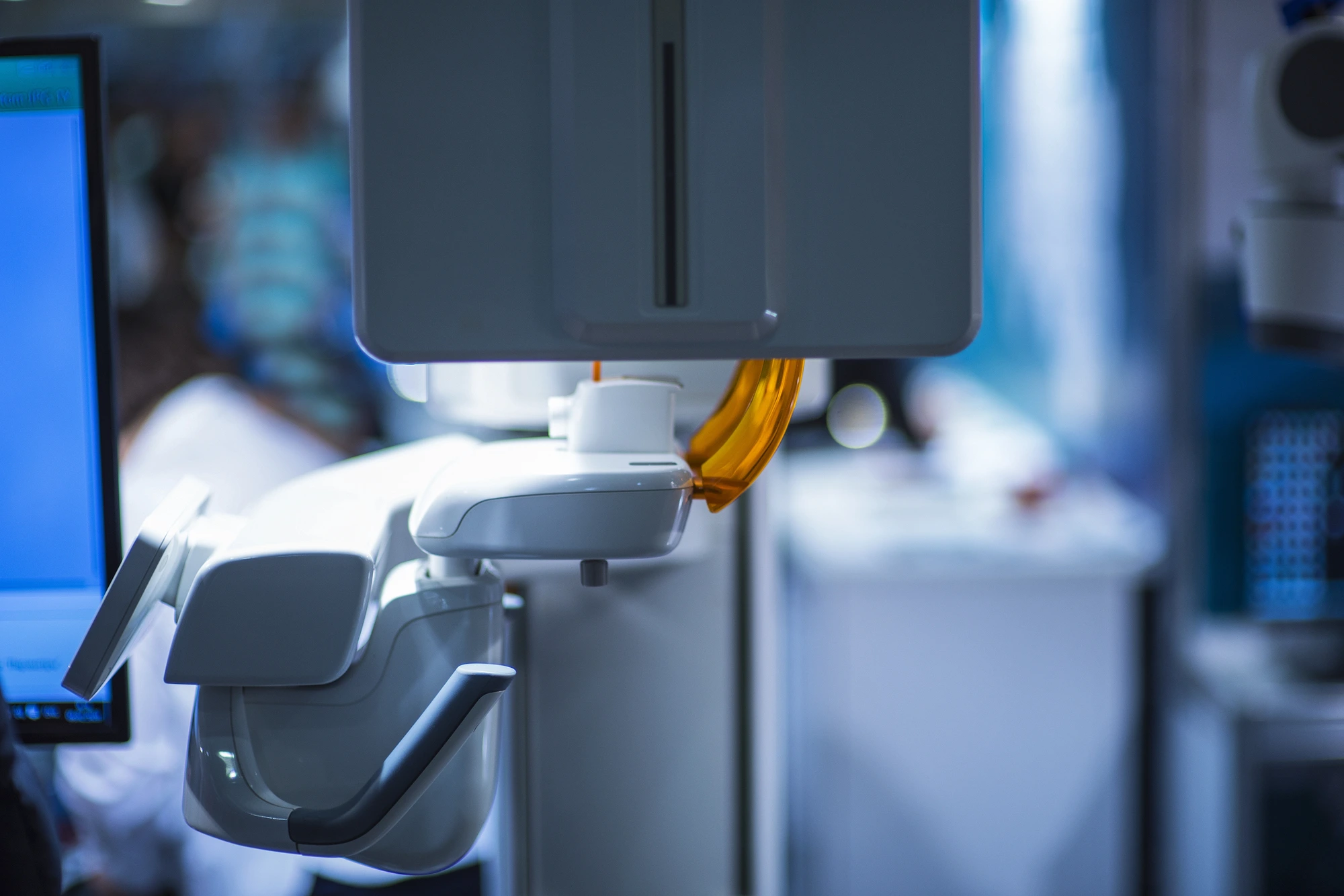
A regular dental X-ray is a flat picture of a three-dimensional tooth, and some root canals hold surprises that a flat image cannot show. When you are planning root canal therapy in Canton, 3D imaging can reveal what a standard X-ray misses, in the right cases. Understanding what 3D cone beam imaging does can help you ask better questions before treatment begins.
Where a Regular X-Ray Falls Short
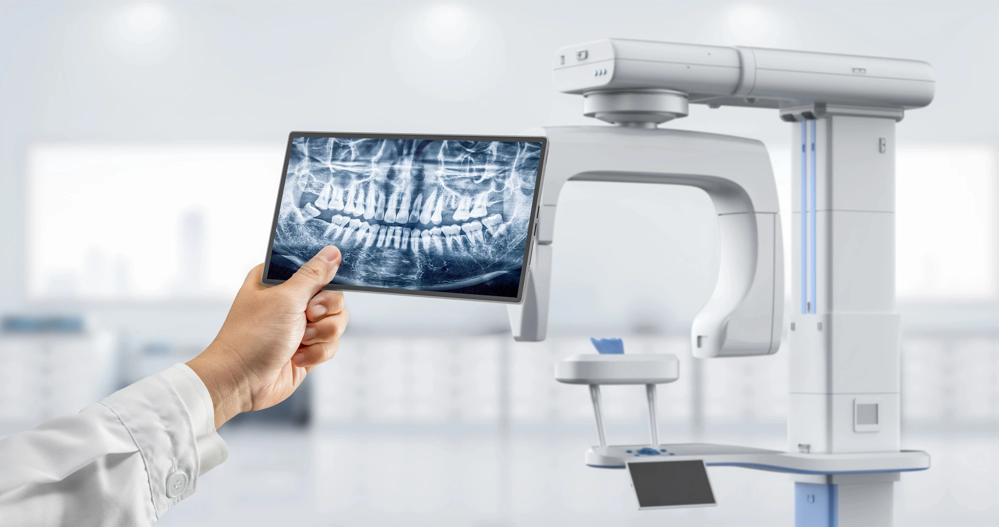
Standard dental X-rays are the first choice for evaluating a tooth. Still, they flatten roots, bone, and surrounding structures into a single image, so overlapping detail can be hidden. The American Association of Endodontists and the American Academy of Oral and Maxillofacial Radiology note in their joint position statement that complex anatomy can make flat images difficult to interpret.
The same statement recommends limited field-of-view 3D imaging for the initial treatment of teeth with potential extra canals and suspected complex anatomy, such as premolars and molars. It also cites a study in which conventional X-rays detected lesions at the root tip in 12% of roots, compared with 25% on 3D scans.
What Can 3D Imaging Show?
A 3D scan can help your dentist see:
- Extra or hidden canals that could be missed during treatment
- Unusual root shapes or sharp curves
- Infection or bone changes around the root tip
- A cracked or fractured root, when other findings are inconclusive
- The tooth's position relative to nearby structures
With that information, your dentist can map the tooth before treatment begins, so the plan reflects what is actually there.
When a 3D Scan Is Not Needed
Many root canals are planned with standard X-rays alone. The position statement says 3D imaging should not be used routinely for diagnosis or screening without clinical signs and symptoms, and only when the benefits outweigh the risks.
The FDA adds that dental CBCT typically delivers more radiation than conventional dental X-rays and should be performed only when it provides information that other imaging cannot. A limited field of view, which scans only the tooth or area of interest, keeps exposure lower.
How 3D Imaging Fits Into Treatment
When a scan is recommended, the process usually looks like this:
- An exam and a discussion of your symptoms
- Standard X-rays
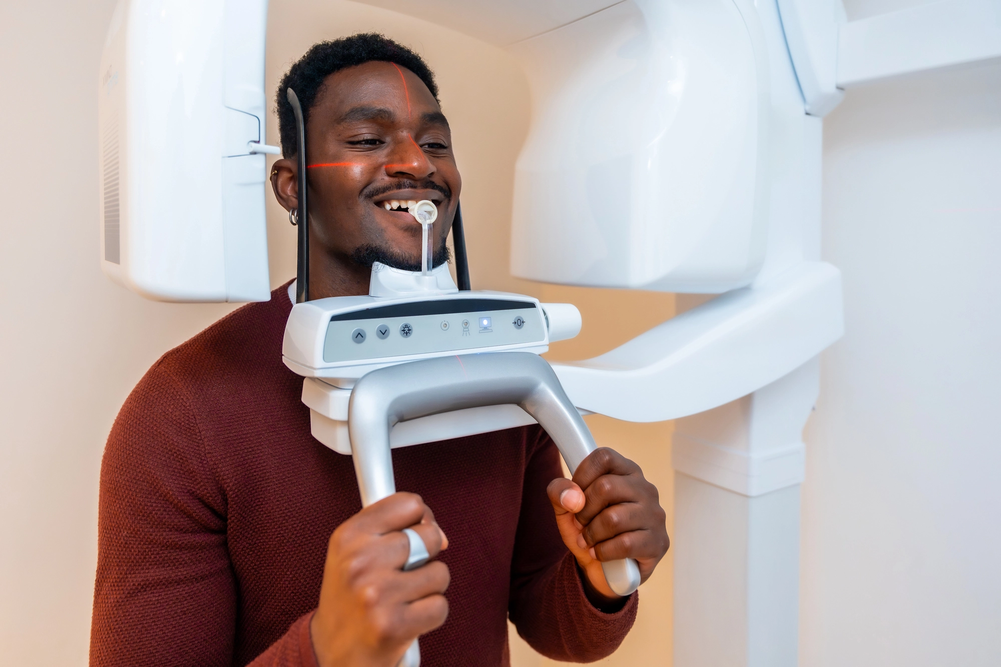
- A 3D scan if your case is complex or the results are unclear
- A treatment plan based on the tooth's actual anatomy
- Treatment with comfort options, including sedation dentistry if you are nervous, and a permanent restoration afterward
Frequently Asked Questions
Does every root canal need a 3D scan?
No. It is used for selected cases, such as complex anatomy or unclear findings.
Is a 3D scan safe?
It delivers more radiation than a standard X-ray, so it is used only when the benefit justifies it, with a limited field of view.
Will the scan make my root canal more complicated?
No. It is a planning tool that helps your dentist prepare.
Need Root Canal Therapy in Canton? Let's See the Whole Picture Before We Treat
Our team at Thomas Dental Group is here to find the source of your pain and plan your care carefully. Schedule your exam or call us at (330) 492-1730 and get lasting dental relief.
Ready to start your journey
Schedule your appointment today and discover the difference quality care makes.

Contact us
We’re ready to help. Reach out with questions or to schedule your visit.